
Body Care
Psoriasis Misdiagnosis: What Else Could It Be?
Jul
Red, itchy, scaly skin seems straightforward until several different conditions arrive wearing nearly identical outfits. Psoriasis is a chronic immune-mediated disease, but its plaques can resemble eczema, fungal infections, dandruff, allergic reactions, and even a few less common disorders. That resemblance can lead to a psoriasis misdiagnosis, especially when the rash appears in an unusual location or does not have the textbook silvery scale.
Getting the name right matters. Psoriasis treatment aims to control an overactive immune response, while ringworm requires antifungal medication and contact dermatitis improves only when the offending substance is identified and avoided. Treating every scaly rash as psoriasis is therefore a bit like using a smoke alarm to repair a toaster: related to the problem, perhaps, but unlikely to solve it.
Why Is Psoriasis Sometimes Misdiagnosed?
Psoriasis can appear almost anywhere, including the scalp, elbows, knees, trunk, skin folds, genitals, palms, soles, and nails. Its appearance also varies by psoriasis type, disease severity, treatment history, and skin tone.
On lighter skin, plaques often look pink or red with white or silvery scale. On darker skin, inflammation may appear violet, deep brown, gray, or darker than the surrounding skin. Scale may be less obvious, and discoloration can remain after inflammation settles. These differences can make image-based self-diagnosis unreliable.
Location creates additional confusion. Thick plaques on the elbows may look classically psoriatic, while smooth redness in the armpit may resemble a yeast infection. Scalp psoriasis can masquerade as stubborn dandruff, and nail psoriasis can be mistaken for nail fungus.
Signs That Make Psoriasis More Likely
A clinician may suspect psoriasis when several of the following features occur together:
- Clearly defined, raised plaques with dry scale
- Similar patches on both sides of the body
- Involvement of the elbows, knees, scalp, lower back, or belly button
- Nail pitting, crumbling, lifting, or discoloration
- A family history of psoriasis
- Repeated flares following illness, stress, skin injury, or certain medications
- Morning joint stiffness, swollen fingers or toes, or other signs of psoriatic arthritis
None of these clues proves the diagnosis on its own. Dermatology is detective work, and the skin does not always cooperate with the witness interview.
Common Conditions Mistaken for Psoriasis
1. Atopic Dermatitis and Other Forms of Eczema
Eczema is one of the most common psoriasis look-alikes. Both conditions can produce inflamed, dry, itchy, cracked, and scaly skin. However, eczema is often itchier and may develop poorly defined borders, swelling, blisters, oozing, or crusting during an active flare.
Atopic dermatitis commonly affects the creases of the elbows and knees, although it can appear elsewhere. Psoriasis more often favors the outer surfaces of the elbows and knees. That distinction is useful, but it is not an unbreakable law. Children may show less typical patterns, and some people genuinely have both psoriasis and eczema.
Another clue is texture. Chronic eczema can become thick and leathery after repeated scratching, while plaque psoriasis tends to form more sharply outlined patches with layered scale. A dermatologist may consider symptoms, location, age of onset, allergy history, family history, and response to previous treatments.
2. Seborrheic Dermatitis
Seborrheic dermatitis affects oil-rich areas such as the scalp, eyebrows, sides of the nose, ears, beard area, eyelids, and upper chest. It commonly produces dandruff and greasy white or yellowish flakes.
Scalp psoriasis often creates thicker, drier scale and may extend beyond the hairline. Seborrheic dermatitis is more likely to have thinner, oilier flakes and to concentrate around the eyebrows, ears, and nose. Unfortunately, skin conditions are under no obligation to respect neat comparison charts.
Some people develop features of both diseases, a pattern often called sebopsoriasis. When the clinical picture remains unclear, a dermatologist may use treatment response or, less commonly, a skin biopsy to clarify the diagnosis.
3. Ringworm and Other Fungal Infections
Ringworm is not a worm. It is a contagious fungal infection known medically as tinea. On the body, it often forms an itchy, circular or oval patch with a raised, scaly outer edge. The center may become clearer as the border expands.
Psoriasis can also create round plaques, so shape alone is not enough. Ringworm may be more likely when a patch expands outward, occurs after contact with an infected person or animal, or appears alongside athlete’s foot or jock itch.
Scalp ringworm can cause scaling, broken hairs, tender areas, and patches of hair loss, particularly in children. These findings deserve prompt evaluation because scalp ringworm usually requires an oral antifungal medication rather than an ordinary skin cream.
A clinician can gently scrape the affected area and examine the sample for fungus. This simple test can prevent months of incorrect treatment. Steroid creams may temporarily reduce redness but can allow a fungal infection to spread and change its appearance, making the eventual diagnosis more difficult.
4. Allergic or Irritant Contact Dermatitis
Contact dermatitis develops when the skin reacts to something it has touched. Irritant contact dermatitis can result from soaps, detergents, solvents, repeated handwashing, disinfectants, or other harsh substances. Allergic contact dermatitis can be triggered by fragrance, nickel, preservatives, hair dye, rubber chemicals, adhesives, cosmetics, or topical medications.
The rash may burn, sting, itch intensely, swell, crack, or blister. Distribution offers an important clue. A rash beneath a watchband, around a belt buckle, under a fitness tracker, or exactly where a cosmetic is applied points toward contact dermatitis rather than psoriasis.
Reactions can be delayed, so the responsible product may not seem suspicious at first. When an allergy is possible, dermatologists may perform patch testing. Small amounts of potential allergens are placed on the back and checked for delayed reactions.
5. Pityriasis Rosea
Pityriasis rosea often begins with one large oval, scaly area called a herald patch. Days or weeks later, smaller spots may spread across the trunk. On the back, the pattern can follow the ribs and resemble drooping branches on a Christmas tree.
Because the smaller lesions can look like guttate psoriasis, confusion is understandable. Pityriasis rosea usually resolves on its own within several weeks and generally does not keep returning. Psoriasis is chronic and tends to cycle through flares and quieter periods.
Atypical pityriasis rosea may lack a noticeable herald patch or may appear in unexpected locations. A clinician may order additional testing when the pattern could represent a fungal infection, medication reaction, or secondary syphilis.
6. Inverse Psoriasis, Intertrigo, and Yeast Infection
Inverse psoriasis affects skin folds, including the armpits, groin, buttock crease, and areas beneath the breasts. Friction and moisture usually reduce the familiar dry scale, leaving smooth, shiny, inflamed patches.
That appearance overlaps with intertrigo, a general term for inflammation where skin rubs against skin. Yeast or bacterial growth can complicate intertrigo. A Candida yeast infection may have small separate bumps or pustules near the main rash, sometimes called satellite lesions.
Inverse psoriasis may also appear in more than one fold and coexist with psoriasis on the scalp, nails, elbows, or knees. A skin scraping, culture, examination under special lighting, or biopsy may be needed when the cause remains uncertain.
7. Lichen Planus
Lichen planus is an inflammatory disorder that can affect the skin, mouth, scalp, genitals, and nails. Classic skin lesions are firm, flat-topped, itchy bumps that may appear red, purple, brown, or gray. Fine white lines can sometimes be seen across the surface.
Thick lichen planus plaques on the lower legs may resemble psoriasis. Oral sores, lacy white patches inside the cheeks, scarring hair loss, or distinctive nail changes may steer the diagnosis toward lichen planus. A skin biopsy is sometimes used for confirmation.
8. Cutaneous Lupus
Lupus can produce scaly, ring-shaped, or disk-shaped patches that resemble psoriasis. Cutaneous lupus often favors sun-exposed areas, and some forms can leave scarring, pigment changes, or permanent hair loss when the scalp is affected.
Photosensitivity, mouth sores, fatigue, joint symptoms, or other systemic complaints may increase suspicion, although skin-limited lupus can occur without obvious internal symptoms. Diagnosis may involve a biopsy, blood tests, and review of medications and sun-related patterns.
9. Cutaneous T-Cell Lymphoma
Cutaneous T-cell lymphoma, or CTCL, is a rare group of cancers involving immune cells in the skin. Its most common form, mycosis fungoides, can initially look like eczema or psoriasis, sometimes for years.
Possible warning signs include persistent patches that do not behave as expected, lesions in areas usually covered by clothing, variation in shape or color, progressive thickening, or poor response to appropriate therapy. These features do not automatically mean cancer; common inflammatory rashes remain far more likely.
When CTCL is suspected, a dermatologist may perform one or more biopsies. Early disease can be difficult to distinguish under the microscope, so repeat sampling and ongoing follow-up are occasionally necessary.
10. Secondary Syphilis
Secondary syphilis can cause a widespread rash that occasionally resembles guttate or plaque psoriasis. Involvement of the palms and soles, swollen lymph nodes, fever, sore throat, patchy hair loss, or sores in the mouth or genital area may provide additional clues.
Because symptoms vary widely, diagnosis depends on appropriate blood testing and evaluation of sexual exposure history. Syphilis is treatable, but missing it can allow the infection to progress and creates a risk of transmission to others.
11. Pityriasis Rubra Pilaris
Pityriasis rubra pilaris is an uncommon inflammatory disease that may cause orange-red scaly plaques, rough bumps centered on hair follicles, and thickening of the palms and soles. Small islands of unaffected skin inside larger inflamed areas are a useful clue.
The condition can resemble extensive psoriasis closely enough that a biopsy and expert dermatology review may be needed. Its rarity also means it may not be considered during an initial primary care visit.
12. Skin Cancer or Precancerous Growths
A rough, persistent, scaly spot may sometimes be actinic keratosis, squamous cell carcinoma, or another skin growth rather than psoriasis. Concern rises when a lesion is solitary, steadily enlarges, bleeds, becomes tender, repeatedly crusts, or fails to heal.
Psoriasis commonly produces multiple recurring plaques, but a new growth should not be automatically assigned to an existing diagnosis. A dermatologist can examine the spot with magnification and perform a biopsy when necessary.
How Doctors Distinguish Psoriasis From Similar Conditions
Medical History
A useful evaluation starts with questions about when the rash began, how quickly it changed, whether it itches or hurts, and which treatments have helped or failed. Clinicians may also ask about recent infections, new medications, travel, pets, occupational exposures, personal care products, family history, and joint symptoms.
Photographs from the beginning of a flare can be surprisingly helpful. By appointment day, a rash may have faded, been scratched, or been altered by several brave experiments from the bathroom cabinet.
Full Skin, Scalp, and Nail Examination
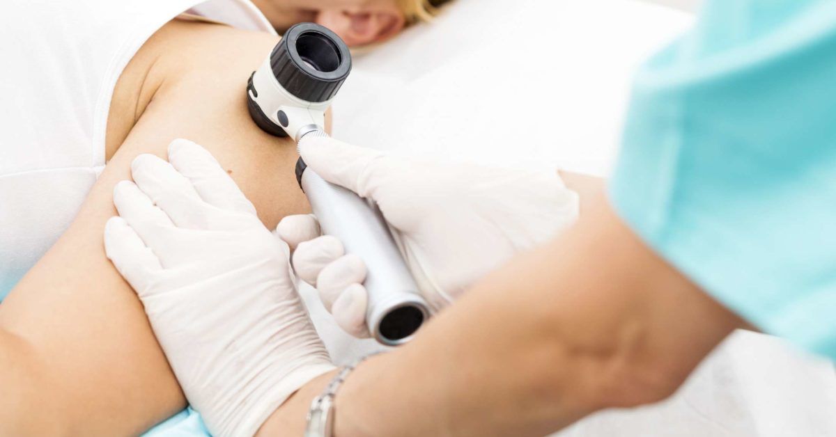
A dermatologist may inspect areas beyond the obvious rash. Nail pits, plaques hidden behind the ears, scalp scale, or inflammation in the belly button can support psoriasis. Broken hairs may suggest scalp ringworm, while mouth lesions can point toward lichen planus, lupus, or an infection.
Examining multiple sites also helps determine whether two conditions are present. A person can have psoriasis and contact dermatitis, psoriasis and a fungal infection, or psoriasis and seborrheic dermatitis at the same time.
Skin Scraping, Culture, and Other Laboratory Tests
When fungus is possible, a clinician may scrape scale from the active edge of a lesion and examine it under a microscope. Fungal cultures or other laboratory methods may be used when the initial result is uncertain.
Blood tests are not routinely needed to diagnose ordinary plaque psoriasis, but they may help investigate lupus, syphilis, infection, medication effects, or other systemic disorders. Patch testing is useful when allergic contact dermatitis is suspected.
Skin Biopsy
Most typical cases of psoriasis can be diagnosed by appearance and history. A biopsy becomes useful when lesions are unusual, treatment repeatedly fails, or the clinician needs to exclude conditions such as lichen planus, lupus, skin cancer, or CTCL.
During a biopsy, the area is numbed and a small sample is removed for microscopic examination. The procedure is generally quick, although the final interpretation must be combined with the clinical picture. Skin pathology is powerful, but it is not a fortune-telling crystal.
Signs That a Psoriasis Diagnosis Should Be Reconsidered
Request a follow-up appointment or dermatology referral when:
- The rash spreads despite correctly used treatment
- Prescribed psoriasis therapy causes unexpected worsening
- A circular patch continues expanding outward
- Only one lesion persists, grows, crusts, or bleeds
- The rash follows the exact shape of clothing, jewelry, or a product application
- There is patchy hair loss or numerous broken hairs
- New mouth sores, fever, swollen lymph nodes, or significant fatigue appear
- The palms and soles develop an unexplained rash
- The diagnosis has changed repeatedly without testing
- You develop joint swelling, prolonged morning stiffness, or swollen fingers or toes
Urgent medical care is appropriate for rapidly spreading redness, extensive skin peeling, widespread pustules, severe pain, fever, facial swelling, trouble breathing, signs of infection, or a rash involving the eyes or mouth. Severe psoriasis and serious medication reactions can affect more than the skin.
Questions to Ask at a Dermatology Appointment
- Which findings support psoriasis rather than eczema or fungus?
- Could two skin conditions be present at the same time?
- Would a fungal scraping, patch test, culture, or biopsy be useful?
- How long should this treatment take to show improvement?
- What changes would mean I should return sooner?
- Do my nail or joint symptoms affect the diagnosis?
- Should I photograph the rash between appointments?
Bring a complete list of medications, supplements, creams, shampoos, and home remedies. Include products that did not work. A failed treatment is not wasted history; it is diagnostic information.
Experiences and Lessons From a Possible Psoriasis Misdiagnosis
The following composite experiences illustrate common diagnostic journeys. They are not accounts of specific patients, but they reflect situations that frequently occur when similar-looking rashes overlap.
The “Dandruff” That Would Not Stay on the Scalp
Someone may spend months rotating through dandruff shampoos because flakes keep returning. The first products help slightly, but thick scale eventually extends behind the ears and beyond the hairline. A closer examination reveals small plaques on the elbows and pitting in several fingernails. What initially looked like ordinary dandruff is diagnosed as scalp psoriasis.
The lesson is not that every flaky scalp means psoriasis. Dandruff is extremely common. The useful clue is the complete pattern: thicker scale, extension outside the scalp, nail changes, and plaques elsewhere. A diagnosis becomes clearer when the clinician examines more than the area named in the appointment request.
The “Psoriasis” That Quietly Expanded
Another person develops one itchy, round patch and assumes it is psoriasis because a relative has the disease. A steroid cream reduces the redness for a few days, but the patch grows wider and develops an irregular scaly edge. Testing later confirms ringworm.
This experience demonstrates why borrowed prescriptions and mystery combination creams can complicate matters. Steroids may alter a fungal rash and allow it to spread. The resulting lesion can look less like classic ringworm and more like an unusual inflammatory disorder, sending the diagnostic process on an unnecessary scenic route.
The Rash That Followed a Fitness Tracker
A person with known psoriasis notices an itchy patch beneath a watchband and assumes it is a new plaque. Stronger psoriasis medication produces little improvement. The sharp rectangular distribution eventually prompts patch testing, which identifies an allergy associated with a material in the band.
Having psoriasis does not make someone immune to other skin problems. Once a diagnosis enters the medical record, it can become the default explanation for every future rash. Patients can help prevent this anchoring effect by mentioning new products, jewelry, work exposures, adhesives, gloves, and changes in skin-care routines.
The Skin-Fold Rash Treated Three Different Ways
Inflammation beneath the breasts or in the groin may be labeled yeast infection, intertrigo, eczema, or inverse psoriasis at different visits. This is not always evidence of careless medicine. These conditions can look very similar, and more than one may occur simultaneously.
Useful details include whether the rash is smooth or scaly, whether separate bumps surround it, whether similar inflammation occurs in several folds, and whether psoriasis appears on the scalp or nails. A scraping can identify fungus, while a biopsy may be considered when the rash remains unexplained.
The Diagnosis That Needed a Second Look
Some people follow a psoriasis treatment plan carefully but see no meaningful improvement. They may blame themselves, use medication more aggressively, or jump between restrictive diets and expensive supplements. A better response is to return to the diagnostic question.
Appropriate treatment failure does not automatically prove that the original diagnosis was wrong. Psoriasis can be stubborn, and a medication may simply be unsuitable. Still, persistent failure is a valid reason to reconsider infection, allergy, overlapping disease, incorrect medication use, or a less common condition.
A second opinion can be especially helpful when the rash is atypical, a biopsy has never been performed, or the diagnosis has changed repeatedly. Patients should bring photographs, previous pathology reports, medication names, treatment timelines, and notes describing what happened after each therapy.
The Emotional Side of Being Misdiagnosed
A prolonged skin problem can affect sleep, clothing choices, work, relationships, and self-confidence. Being told that a visible rash is “just stress” can feel dismissive, even though stress may worsen many inflammatory conditions. Conversely, receiving a frightening list of rare possibilities from an online search can turn an ordinary Tuesday into a medical thriller.
The most productive approach sits between those extremes. Track symptoms, seek qualified care, ask how the diagnosis was reached, and request testing when the clinical pattern remains uncertain. A good dermatologist should be willing to explain both the leading diagnosis and the important alternatives.
Most psoriasis look-alikes are manageable once they are correctly identified. The goal is not to become suspicious of every diagnosis. It is to recognize when the rash is behaving differently from the expected script and deserves another examination.
Conclusion
Psoriasis misdiagnosis is possible because many inflammatory, infectious, allergic, autoimmune, and neoplastic skin conditions can produce scaly plaques. Eczema, seborrheic dermatitis, ringworm, contact dermatitis, pityriasis rosea, inverse psoriasis look-alikes, lichen planus, cutaneous lupus, and several rarer conditions belong on the differential diagnosis.
Location, border shape, scale texture, nail findings, exposure history, systemic symptoms, and treatment response all provide clues. When those clues do not form a convincing picture, a fungal scraping, patch test, blood test, culture, or skin biopsy may supply the missing evidence.
The key takeaway is simple: a rash that fails to improve is not asking for increasingly random creams. It is asking for a fresh look.
SEO Information

